Diastasis recti surgery: before and after, explained
This page is a reading guide for people with diastasis recti who are looking at before-and-after photos, here or anywhere else, and want to know what to look for.
A before-and-after image of diastasis recti repair shows how the profile of the abdomen changes once the two rectus muscles are brought back to the midline: less forward bulging when standing, a flatter waistline, a navel that sits differently and a scar whose length depends on the technique. It cannot show core strength or how symptoms have changed. The documented diastasis cases operated on by Dr. Federico Fiori are published, with consent, in the diastasis gallery on federicofiori.com.
What changes in a diastasis recti before and after?
Diastasis recti is a widening of the linea alba, the band of connective tissue between the two rectus muscles. In the 'before' image it usually shows as a bulge along the midline, most visible when standing or straining, sometimes together with an umbilical hernia. In the 'after' image the midline has been repaired: the profile is flatter, the bulge on straining is reduced and the waistline is better defined.
How is a diastasis measured before it is photographed?
A photograph shows shape; the diastasis itself is a width. Before any image is compared, the gap is located along the midline and measured, first by hand and then with a caliper and ultrasound.
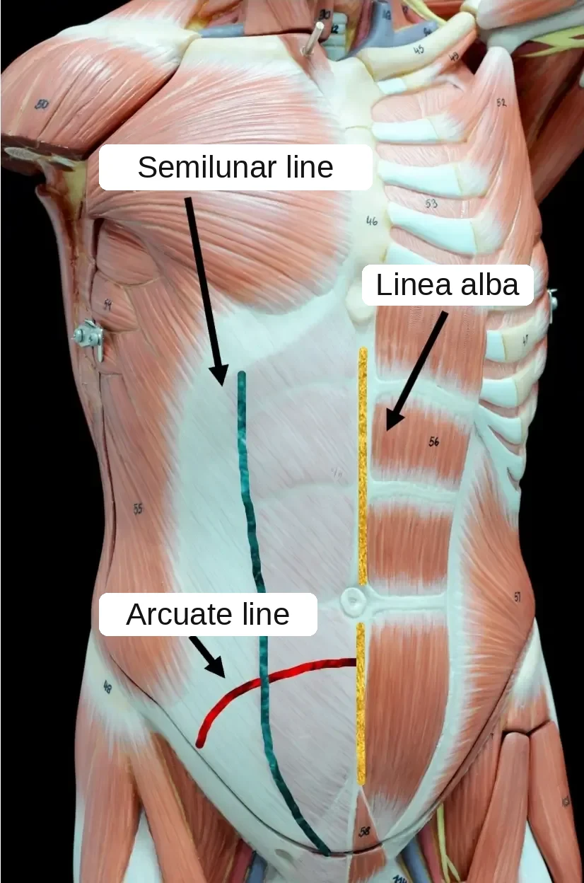
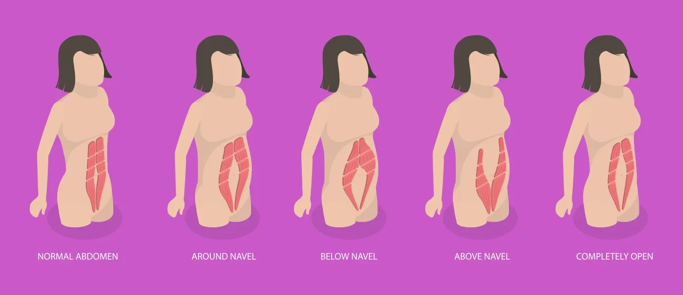
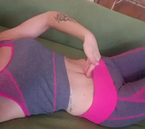
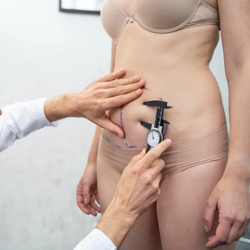
In 150 women who had never been pregnant, the linea alba was up to 15 mm wide at the xiphoid, 22 mm 3 cm above the navel and 16 mm 2 cm below it (Beer GM et al., Clin Anat 2009; DOI 10.1002/ca.20836). Wider values suggest a diastasis; the details are in how rectus diastasis is measured and when it becomes significant.
How does the technique change what you see?
- Minimally invasive repair, such as TESAR: a few short incisions and no removal of skin, so the change is mainly in the profile and in the midline bulge, while loose skin stays as it was;
- Abdominoplasty with repair of the midline: excess skin and fat below the navel are removed, so the change in shape is larger, with a low horizontal scar and a repositioned navel;
- Minimally invasive lipoabdominoplasty (MILA): the midline repair is combined with treatment of fat and skin, for people whose diastasis comes with some excess tissue.
How the technique for diastasis recti is chosen
When is the result of diastasis repair judged?
Not in the first weeks. Swelling along the repaired midline and around the navel takes months to settle, and scars keep changing in color and thickness for a year or more. The images worth comparing are those taken from about six months onward, in the same position as the 'before'.
What a photo of the abdomen cannot tell you
Diastasis recti is not only about shape. Back pain, a feeling of a weak core, pelvic floor symptoms and digestive discomfort are among the reasons people seek repair, and none of them shows in a photograph. Whether they improve is assessed at the follow-up visits, not in pictures.
Whether the change in shape alone is a reason to operate is discussed in rectus diastasis: aesthetic or functional problem?
The symptoms of diastasis recti
Why are the documented cases kept on federicofiori.com?
Clinical photographs are health data. So that each image is published only once, with its consent and its details of technique and follow-up, the diastasis gallery is kept in one place, the surgeon's own site. This page explains how to read it.
Who may use these images?
The images on this site are © Dr. Federico Fiori. Clinical photographs are health data: they are published only with the patient's specific written consent, which can be withdrawn, and they may not be copied or republished without permission. Requests to use an image go through the contact page.
Before and after a second operation, see also a second opinion after abdominoplasty: what can still be corrected.
Before you look at any diastasis photo
Check three things first: whether the 'after' image is taken standing and in profile, like the 'before'; how long after surgery it was taken; and which technique was used. A photo from the first weeks still shows swelling, and an abdomen photographed from a different angle can look flatter than it is.
The diastasis photographs are published only once, on the surgeon's own site, each with the specific written consent of the patient and without recognizable faces. Open the gallery with the checklist above in mind: same position, time since surgery, technique. See the diastasis cases
Questions about diastasis recti before and after
- Will my abdomen look like the diastasis cases in the gallery?
- Not necessarily. Each image shows one person's diastasis, skin and starting weight. What can be achieved in your case depends on the width and length of the gap, the amount of loose skin and whether there is an umbilical hernia, and it is established at the consultation.
- Does a flat abdomen in the 'after' photo mean the diastasis is fixed?
- A flatter profile is a good sign, but the repair itself is checked by examination and, when needed, ultrasound: a photo shows the surface, not the midline.
- Why do some 'after' photos still show a small bulge?
- Because the shape also depends on fat inside the abdomen, on posture and on the time since surgery. Repairing the midline does not remove visceral fat, and early swelling can look like a bulge for several months.
- Is there a visible scar after minimally invasive diastasis repair?
- The operation is done through a few short incisions rather than one long one, so the visible scars are limited. After an abdominoplasty the scar is longer and runs low across the abdomen.
- Where can I see the documented diastasis cases?
- In the diastasis gallery on federicofiori.com, where each case states the technique and the time since surgery.
Updated: 2026-10-18 · Content by Dr. Federico Fiori