What is diastasis recti?
Diastasis recti is caused by the thinning and widening of the linea alba, the connective tissue that joins the two rectus abdominis muscles down the midline, so the two muscles drift apart. It reduces the abdominal wall's ability to contain and stabilize the trunk, and is a functional problem — not only a cosmetic one.
Diastasis recti is a condition in which the linea alba — the connective band that joins the two rectus abdominis muscles down the midline — loses its normal integrity and tension. When it gives way and widens, the two muscles separate by a gap that is not normally present, and the whole abdominal core starts to work less efficiently. It is not a life-threatening condition, but it can cause genuinely disabling symptoms and, in some people, a true change in the shape of the abdomen.
What exactly is diastasis recti?
Diastasis recti (rectus diastasis) is defined as a loss of integrity and tension of the linea alba, the fibrous seam that unites the right and left rectus abdominis muscles along the midline. When the linea alba stretches, becomes lax and widens, the distance between the two muscles increases, they move laterally, and they lose their normal characteristics and function — with consequences for the entire abdominal core.
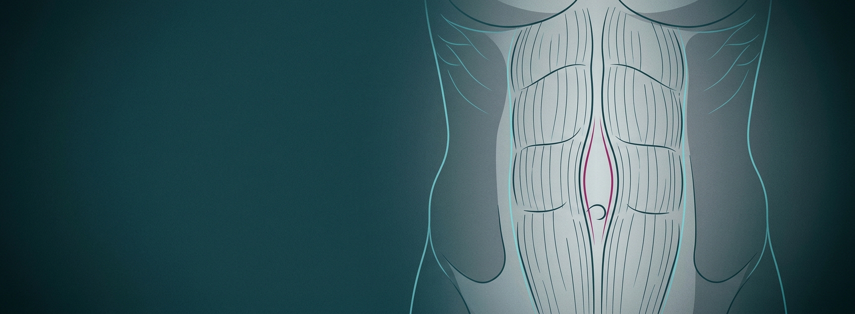
A seam under tension
Think of the linea alba as a central seam that keeps the two halves of the front of the abdomen together and lets them work as one. When the seam loosens, the wall can no longer tension itself properly.
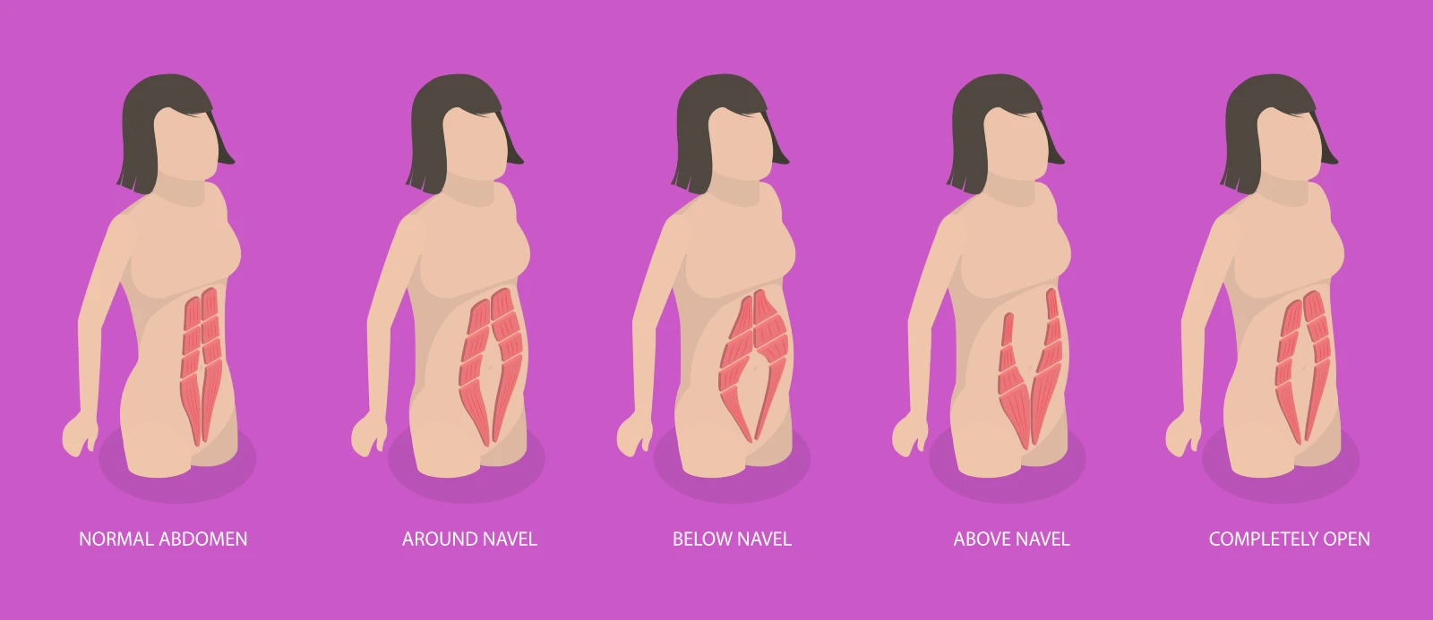
Muscles pushed apart
As the linea alba loses tension, the two rectus muscles separate and the wall stops working as one.
How is the abdominal wall built?
The anterior abdominal wall is layered. From the outside in: skin, subcutaneous tissue, superficial fascia, the muscles with their aponeuroses, and the transversalis fascia. The muscles are paired and symmetrical — the rectus abdominis, the external and internal obliques, the transversus abdominis and the pyramidalis. Blood vessels, nerves and reference structures such as the linea alba, the arcuate line and the semilunar line complete the picture.
The right and left rectus muscles meet on the midline through the linea alba. A deeper walk-through of these layers is on the abdominal core anatomy page.
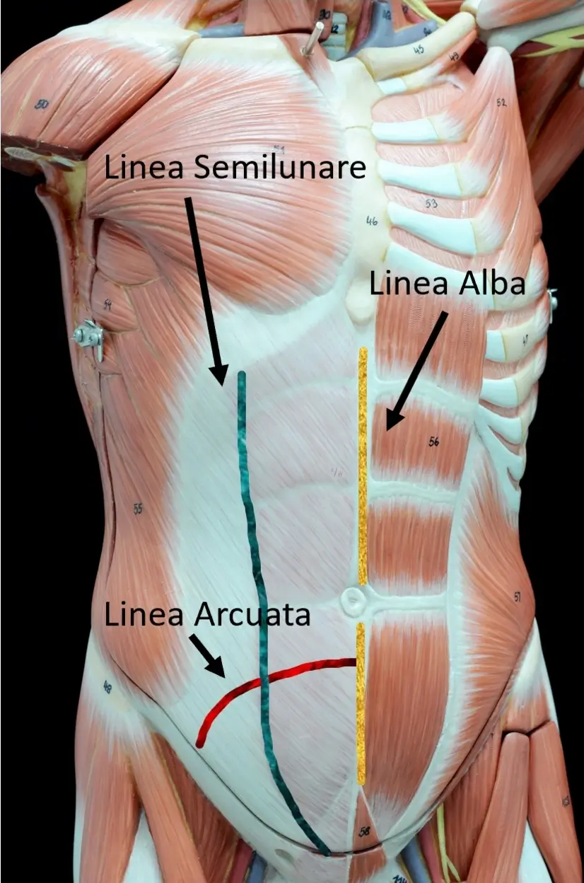
A layered wall
Skin, fat, fascia, muscle and their aponeuroses stack in layers, joined on the midline by the linea alba.
Where along the midline can it occur?
A diastasis does not always look the same. It may involve only the supra- and peri-umbilical portion of the midline (above and around the navel), only the umbilical and infra-umbilical portion (at and below the navel), or the entire midline from the breastbone to the pubis.
What do you feel and what do you see?
Beyond the symptoms you feel, diastasis often produces something you can see. Standing, the abdomen may look prominent — sometimes resembling a mid- or late-pregnancy shape. On effort or coughing, a ridge can appear along the midline, which patients often call a “fin.”
Two components of shape
The look of a standing abdomen is the sum of (1) the wall itself — fascia and muscle, which may be toned, hypotrophic or under-functioning — and (2) the skin and fat above it, which may be scant, moderate or abundant. These combine in many ways, which is why treatment is never one-size-fits-all.
How does diastasis recti differ from an umbilical hernia?
Diastasis is frequently associated with an umbilical hernia — a protrusion of tissue from inside the abdominal cavity through a defect in the wall at the navel. The two are different: a diastasis is caused by a widening of the seam between healthy muscles, whereas a hernia is a true hole through which contents can push. Because they can coexist and change the treatment plan, any firm or tender midline lump deserves assessment.
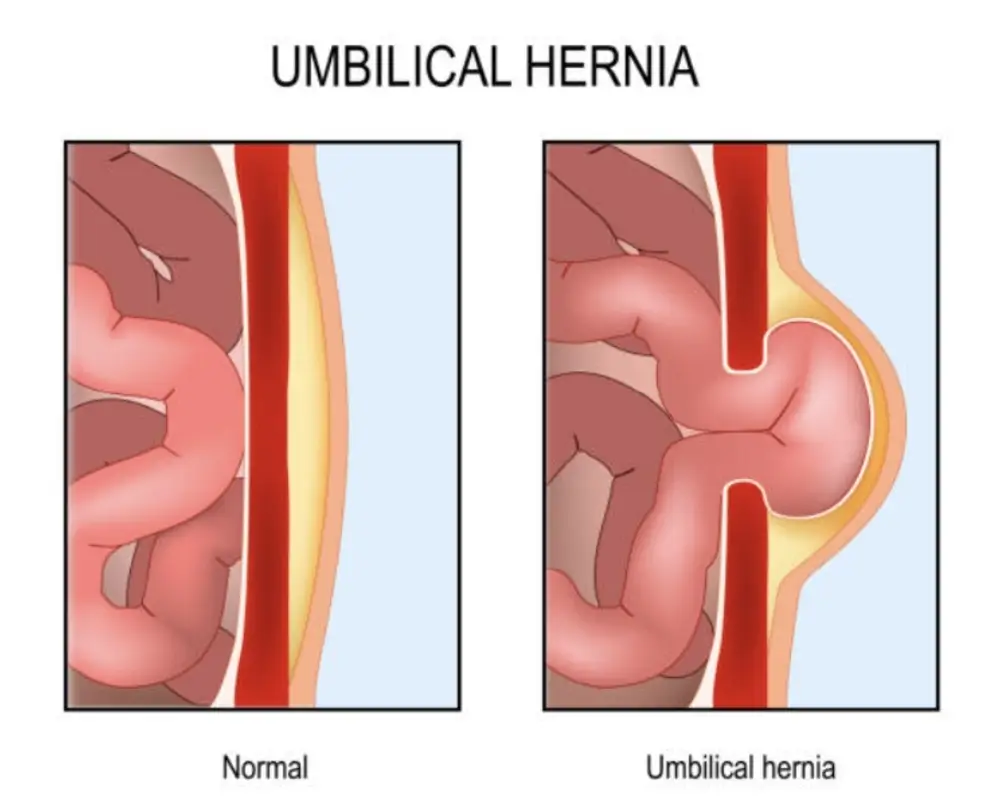
Often a hernia too
An umbilical hernia lets tissue push through a defect at the navel and frequently coexists with a diastasis.
What is diastasis recti in simple terms?
It is a loss of tension and integrity of the linea alba, the connective seam joining the two rectus abdominis muscles. The muscles separate by a gap that is not normally there, and the abdominal wall works less efficiently.
Is diastasis recti dangerous?
It is not a life-threatening condition and rarely needs urgent treatment. However, it can cause disabling symptoms and a visible change in the shape of the abdomen, so it should be assessed and managed properly.
Is diastasis recti the same as a hernia?
No. A diastasis is caused by a widening of the linea alba between intact muscles, while an umbilical hernia is a defect through which tissue can protrude. They often coexist, and an ultrasound tells them apart.
Does the size of the gap predict how bad the symptoms are?
No. The symptoms of diastasis are non-specific and are not proportional to the width of the gap: a narrow diastasis can be symptomatic and a wide one relatively quiet.