Diagnosis and imaging of diastasis recti
Diastasis recti is diagnosed by measuring the inter-recti distance. Ultrasound is the first-line test — dynamic, radiation-free and able to rule out an associated hernia — while CT or MRI are reserved for complex or recurrent cases.
A home self-check raises the suspicion; imaging confirms it. The goal of radiological diagnosis is to document and quantify the distance between the rectus muscles along the whole linea alba and to identify any associated hernia. Ultrasound is the reference first-line test; CT and MRI are second-line, for complex cases.
Why is ultrasound the first-line test?
Ultrasound is the logical first step: it needs no preparation, is rapid, completely non-invasive and uses no ionizing radiation. Crucially it is dynamic — the wall can be assessed standing and during a contraction or Valsalva maneuver, which is especially useful for spotting small hernias. High-frequency linear probes (typically 10–18 MHz) give excellent resolution, usually with the patient lying supine.
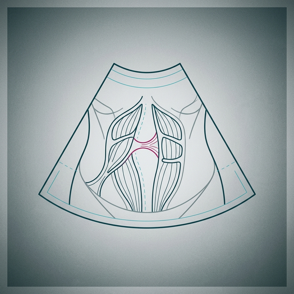
What it shows
It visualizes the linea alba from the xiphoid down to the pubis, measures the width and extent of the gap, and checks its continuity to find fascial defects or hernia openings (often at the umbilical and epigastric levels). It can also assess muscle trophism and the thickness of the subcutaneous layer.
How is the gap measured — and why does the number matter?
The examiner measures the inter-recti distance (IRD) in millimeters, at several levels, at rest and on effort. A diastasis is often diamond-shaped and centered on the navel, where the linea alba is naturally widest. The maximum width is particularly relevant for planning treatment — and in Italy this measurement can influence eligibility for surgery within the National Health Service, which uses a nationally defined threshold.
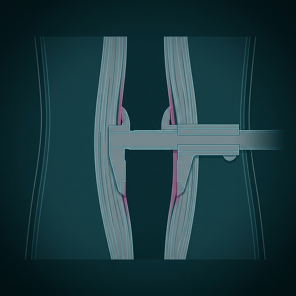
An objective, repeatable number
Unlike fingerbreadths, the ultrasound measurement is reproducible — essential for tracking progress and for planning any surgical repair. Its main limits are that it is operator-dependent and that a probe of ~5–5.5 cm can struggle with very wide diastases.
When are CT and MRI used?
CT and MRI are second-line. They are less operator-dependent and more reproducible, and inherently panoramic, which makes them useful for very large or complex defects such as incisional and other ventral hernias or postoperative collections. Their downsides: they are essentially non-dynamic (they cannot reproduce standing or straining, so small hernias may be missed), they cost more and take longer, and CT uses ionizing radiation — so it should be reserved for when the expected benefit justifies it. The decision to request CT or MRI is a specialist one.
Ultrasound is the sensible first examination when diastasis is suspected, provided it is done in a center experienced in abdominal-wall imaging.
How is diastasis recti diagnosed?
By a clinical examination plus imaging. Ultrasound is the first-line test: it measures the inter-recti distance at rest and on contraction, assesses the linea alba and rules out an associated hernia.
Why is ultrasound preferred over CT or MRI?
Ultrasound is quick, painless, radiation-free and dynamic — it can assess the wall standing and on straining, which helps detect small hernias. CT and MRI are reserved for large, complex or recurrent defects.
Do you need an MRI or CT scan for diastasis recti?
Usually no. Ultrasound is the reference test. CT or MRI is reserved for complex or pre-surgical cases, and the decision is made by the surgeon; CT in particular uses radiation and is used only when justified.
Does the measured width affect whether surgery is covered?
It can. The maximum inter-recti width guides treatment, and in Italy it can influence eligibility for surgery within the National Health Service, which uses a nationally defined threshold.