Surgical treatment of diastasis recti
Surgery for diastasis recti is considered when the gap is wide or long-standing, when symptoms persist despite rehabilitation, or when a hernia is also present. It rebuilds a closed, mechanically effective abdominal wall using open abdominoplasty or minimally invasive techniques such as TESAR, SCOLA, eTEP and robotic repair.
Surgery is the tool that removes the anatomical cause of the symptoms. It rebuilds a closed, mechanically effective abdominal wall by bringing the rectus muscles back to the midline and, when needed, reinforcing it. An important truth first: surgery restores tension and function, but it does not recreate lost muscle tone — it does not, by itself, create a “flat stomach.” The right technique is chosen with a morpho-functional approach.
Does every diastasis need surgery?
No. Surgery is proposed when there are symptoms that can be linked to the diastasis — functional and/or morphological. If there are no relevant symptoms it can be deferred, keeping in mind that the muscle will keep working inefficiently, so symptoms may still appear in future. The gap does not necessarily widen over time, but the muscle hypotrophy tied to an under-functioning abdominal core can appear or worsen.
There is no single operation for everyone. The surgeon must explain the origin of the discomfort and the different options, considering the “diastasis problem” as a whole — wall, muscle, fat and skin together.
What can surgery achieve — and what can it not?
Repairing the wall puts the muscular system back “under tension” and removes the clinical-functional symptoms, but it does not by itself restore lost muscle tone. To be concrete: surgery does not create a “flat stomach,” it rebuilds a closed, functioning wall. Rebuilding the linea alba then makes it possible to restart progressive muscular re-education — ideally with hypopressive gymnastics, which does not specifically load the freshly repaired midline. The more toned the wall is before surgery, the more satisfying the immediate result.
How is the right technique chosen?
Diastasis surgery belongs to the wider field of midline reconstruction (the midline being the linea alba, from sternum to pubis). The indication comes from the problem: there are open techniques and minimally invasive techniques, and each patient should be offered the correct one based not only on symptoms but also on morphology. A patient with major skin excess after big weight loss has a clear indication for laparoplasty with abdominoplasty; a very slim patient with no redundant skin is directed toward a minimally invasive repair. Between these extremes lie all the shades of gray, where expected results are balanced against the risks of each option. Whenever the fascia and muscles are repaired, it is called a laparoplasty — every other label is accessory.
The right technique is chosen with a morpho-functional approach.
Which techniques repair a diastasis, and how do they differ?
The right technique depends on how wide the diastasis is, whether a hernia is also present, how much excess skin there is, and the individual patient’s characteristics. The options below range from open repair with abdominoplasty to minimally invasive and robotic approaches — the choice is made at the consultation, not from a single measurement.
Open surgery and abdominoplasty
The open technique is used when redundant skin and fat must be removed — it is the only approach that delivers a functional repair and the fullest morphological correction. It uses a suprapubic incision (in women, often along an old cesarean scar); the flap is then lifted to the navel, where a first technical decision is made.
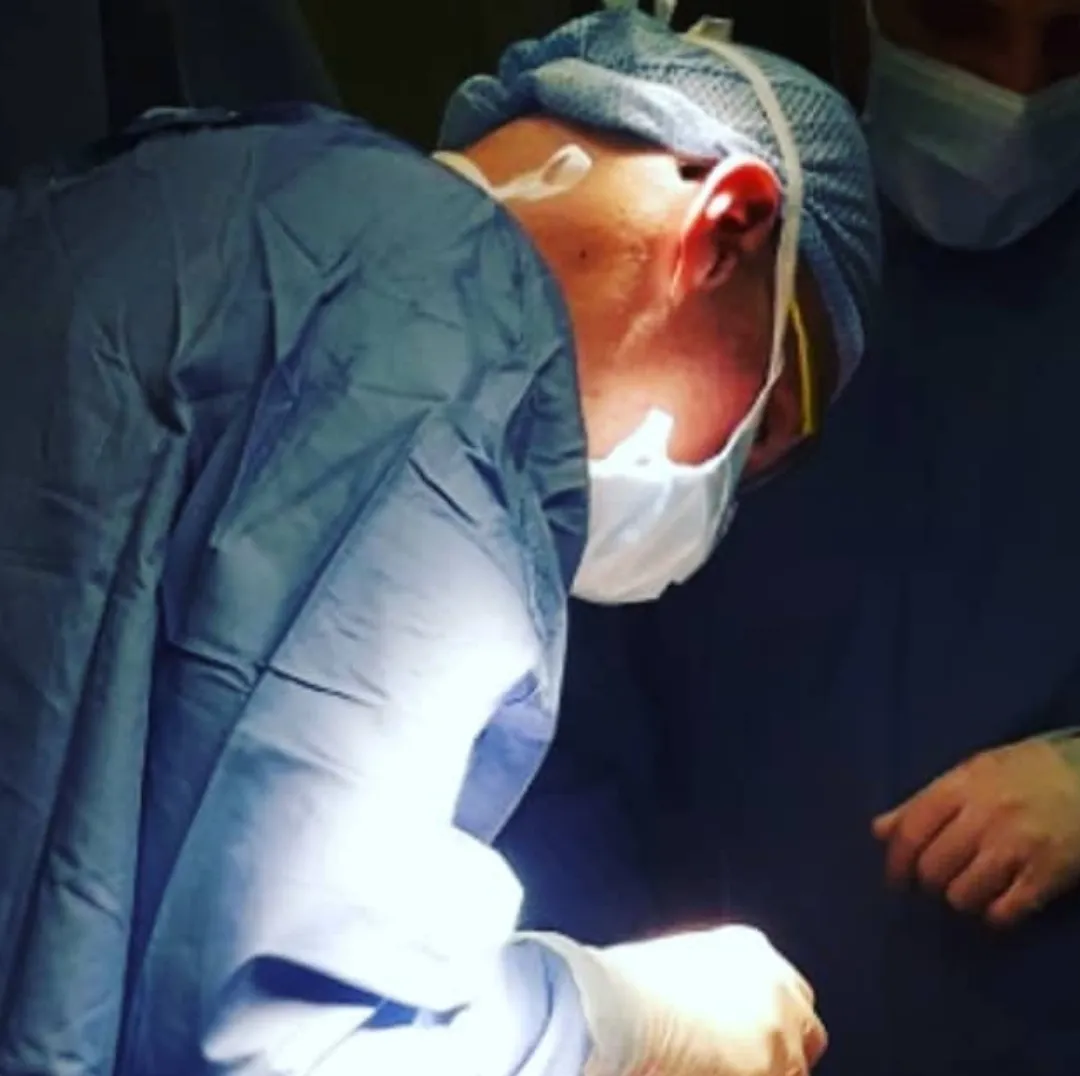
The open route
Open surgery is the only approach that removes excess skin and fat while repairing the wall.
Laparoplasty with mini-abdominoplasty
Chosen when there is little skin-fat excess, or when it is mainly below the navel; not suitable for major excess above or around the navel. A single suprapubic scar, roughly from one iliac spine to the other. The flap is lifted to the navel, the navel is separated from the fascia (revealing any umbilical hernia), dissection continues to the sub-xiphoid region, then the diastasis and muscle trophism are assessed and the wall is repaired with or without a mesh.
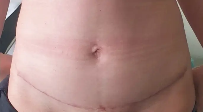
Mini-abdominoplasty
A single suprapubic scar, for limited or mainly lower-abdominal skin excess.
Full abdominoplasty and the inverted-T scar
For major skin excess, especially above the navel, a full abdominoplasty with transposition of the navel is needed. The navel is cut free from the surrounding skin but stays attached deep, in place, while the flap slides down over it. The umbilical hernia is repaired first (a defect up to ~1 cm with 1–2 direct sutures; larger defects with a mesh), then the diastasis is plicated in two segments — sternum-to-navel and navel-to-pubis — over a mesh when indicated, and the obliques may be plicated to reduce the waistline further.
Why does one operation end with a horizontal scar and another with an inverted-T? If the upper flap is long enough, the old navel opening reaches the horizontal suprapubic incision and everything extra is removed with a horizontal scar only. If it is not long enough, a vertical closure is also needed — the inverted-T — whose length varies from a few centimeters to the whole navel-to-pubis distance.
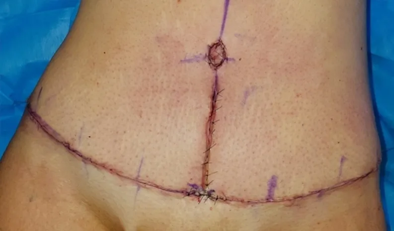
Horizontal or inverted-T
Whether a vertical scar is added depends on how much upper-abdominal skin must be removed.
What is minimally invasive surgery?
Here the surgeon works looking at a screen, creating a working chamber between the layers of the wall with CO₂ and operating through small skin incisions. It can rebuild the wall and repair the diastasis and other defects, restoring the integrity and function of the abdominal core — but it cannot directly remove major excess skin, which only open surgery addresses. Choosing the right indication is therefore essential.
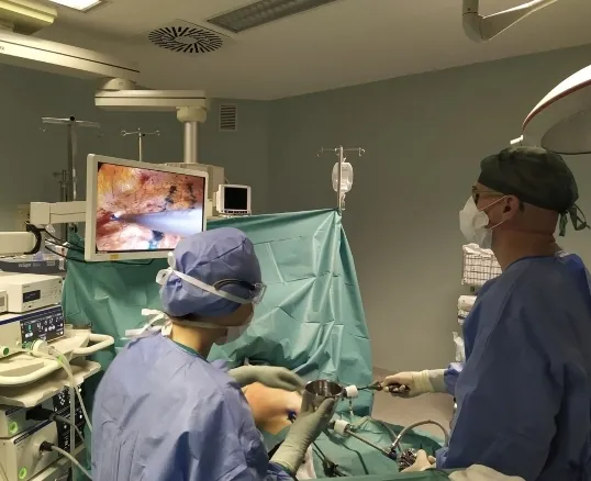
Through small incisions
Minimally invasive surgery rebuilds the wall via small ports, but cannot remove major excess skin.
Techniques are grouped by how they reach the wall — posterior (from behind/beneath the muscles) or anterior (from in front) — and by where the mesh sits: onlay (above the fascia) or sublay (behind the muscle). No technique is universally superior; some patients are simply better suited anatomically to one than another. A plain-language glossary of the acronyms is on the hub: abdominal-wall surgery glossary.
Posterior intraperitoneal techniques: IPOM, LIRA, TAPP
With these the instruments enter the peritoneal cavity, among the organs.
- IPOM (Intraperitoneal Onlay Mesh, more correctly IPUM): a mesh is placed from inside to cover the defect without closing it, so it is not a true diastasis repair. IPOM-Plus adds a sutured closure of the defect before the mesh.
- LIRA (Laparoscopic Intracorporeal Rectus Aponeuroplasty): an evolution of IPOM-Plus — the posterior rectus sheath is incised away from the defect and sutured to rebuild the wall before mesh reinforcement, reducing tension on the central suture line; recent variants place the mesh preperitoneally, outside the cavity.
- TAPP (Transabdominal Preperitoneal): the peritoneum is separated to create a preperitoneal plane; the defect is closed and the mesh placed there, then the peritoneum is closed over it so the mesh stays outside the cavity.
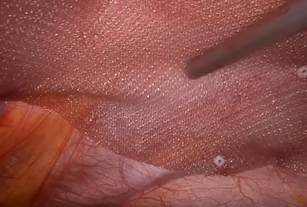
Among the organs
Posterior intraperitoneal techniques work inside the cavity; IPOM-Plus closes the defect before the mesh.
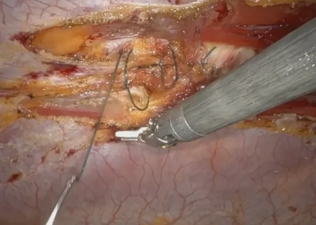
LIRA
The wall is reconstructed and sutured before mesh reinforcement, to reduce tension on the midline.
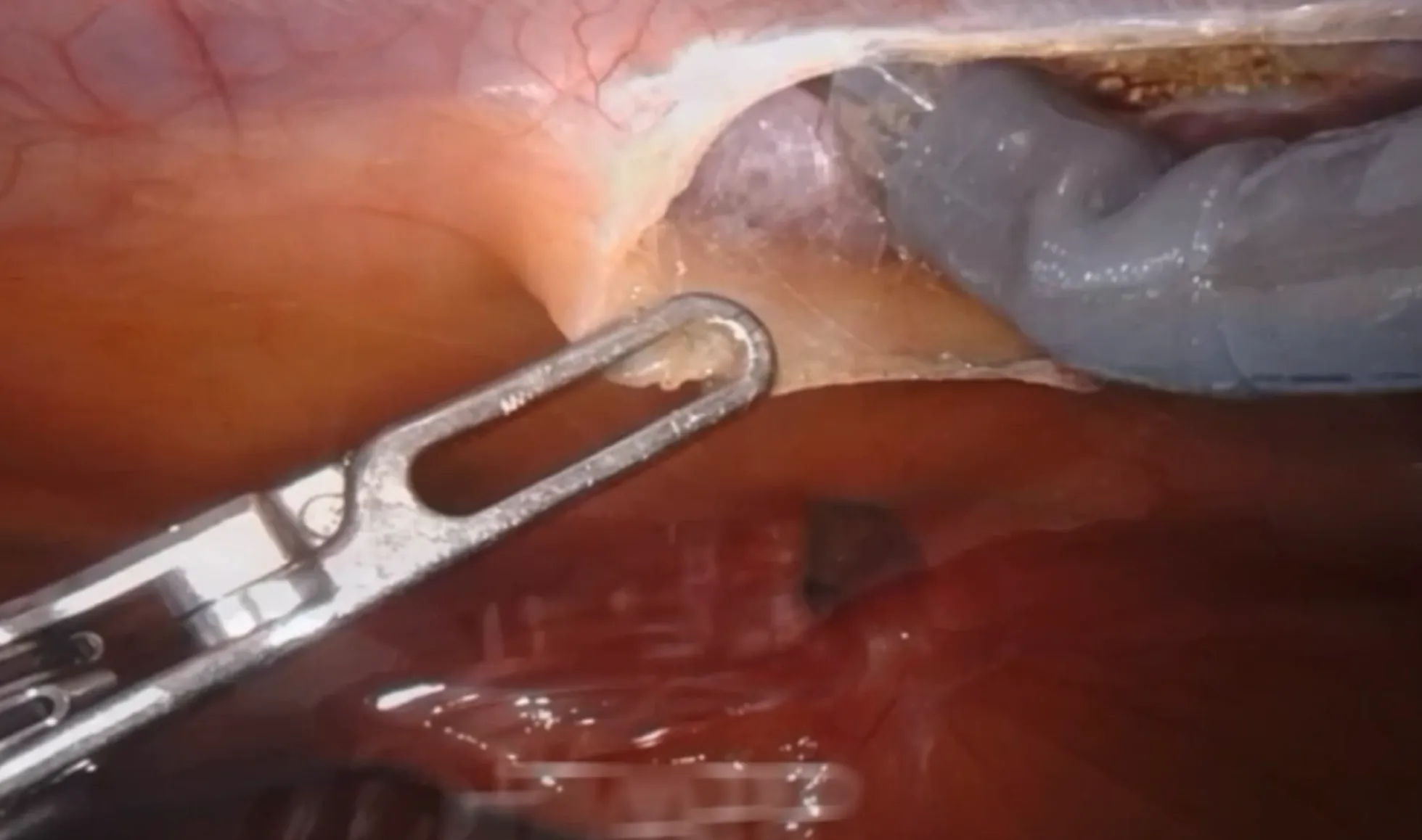
TAPP
A preperitoneal plane is created so the mesh stays outside the abdominal cavity.
Stapler-assisted posterior techniques: THT and MISAR
These “mixed” posterior techniques use a mechanical stapler — which cuts and joins with tiny metal staples — instead of a hand-sewn thread, working in the retromuscular plane under intraperitoneal camera control.
- THT (Trentino Hernia Team): from a single umbilical access the retromuscular plane is entered and the stapler divides and reconstructs the tissues upward and downward under camera control; a retromuscular mesh is then positioned. It made stapled reconstruction reproducible and is spreading rapidly.
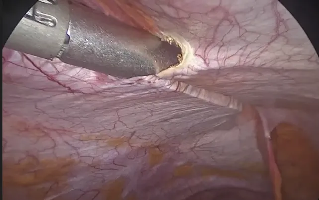
- MISAR (Minimally Invasive Stapled Abdominal Reconstruction): via three suprapubic incisions the posterior sheaths are opened beside the diastasis and stapled together upward, creating space for a retromuscular mesh.
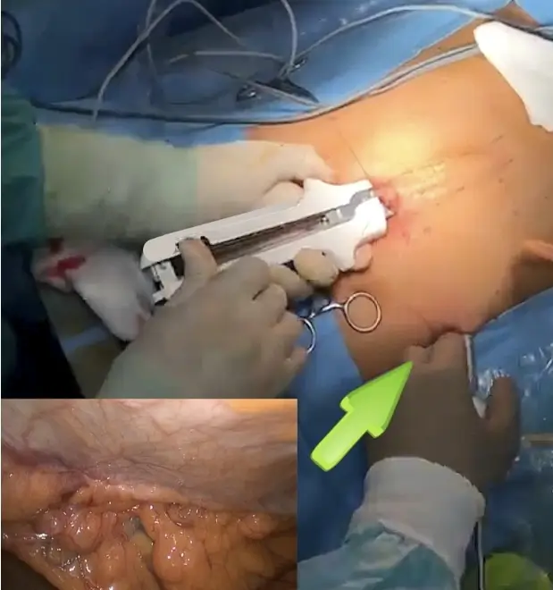
THT
A stapler rebuilds the midline from a single umbilical access, under camera control.
MISAR
Stapled approximation of the muscles through three suprapubic incisions, with a retromuscular mesh.
Posterior extraperitoneal (endoscopic): TEP, eTEP and PeTEP
Endoscopic, not laparoscopic: the instruments never enter the cavity but work in a space the surgeon creates behind the muscles. This is the minimally invasive approach most discussed in recent years, used for the midline as well as groin defects.
- TEP / eTEP (Totally Extraperitoneal / Enhanced-view): the retromuscular space is entered directly and the right and left sides are joined by a “crossover” behind the linea alba, creating one continuous space; the defect is repaired on both planes and a mesh is placed. Today eTEP is often robotic (r-eTEP), which simplifies the otherwise awkward “en reverse” work.
- PeTEP (Preperitoneal eTEP): dissection starts below the arcuate line and progresses between the posterior sheath and the peritoneum; preserving the posterior sheath (rather than incising it) seems to protect against post-operative bulging.
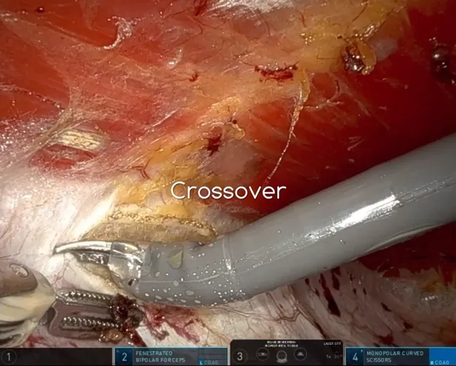
eTEP
A continuous retromuscular space is created behind the muscles, today often robotically.
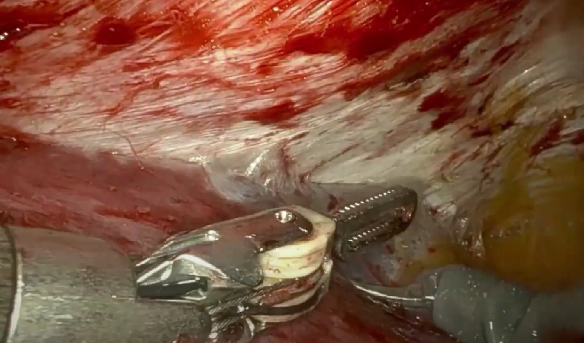
PeTEP
Dissection preserves the posterior rectus sheath, which may protect against post-operative bulging.
Anterior techniques: SCOLA/REPA and TESAR
The anterior route reaches the fascia through small incisions and creates a working chamber in front of the muscles. REPA and SCOLA are the same family: wide prefascial dissection, plication of the fascia and diastasis, and a mesh placed onlay (above the muscles). It is widely used, especially in the version without mesh, with good results.
The onlay position, however, is debated: statistically, wound infections and occurrences (SSI/SSO) are more frequent with onlay meshes. Placing the mesh in a closed space behind the muscles (sublay) reduces those risks. This is exactly the reasoning behind TESAR.
TESAR — developed by Dr. Fiori
TESAR (Totally Endoscopic Sublay Anterior Repair) combines the advantages of the anterior route with a mesh placed sublay, behind the rectus muscles. The prefascial chamber is created as in SCOLA, the umbilical defect is closed, then the medial edge of each rectus sheath is opened and the retromuscular spaces are developed to receive the mesh — while the pathological linea alba stays intact on the floor of the chamber. The mesh reinforces the whole midline without ever opening the posterior plane (except for the small umbilical passage). Dr. Fiori first used it in 2018 and published it in 2019 (J Laparoendosc Adv Surg Tech).
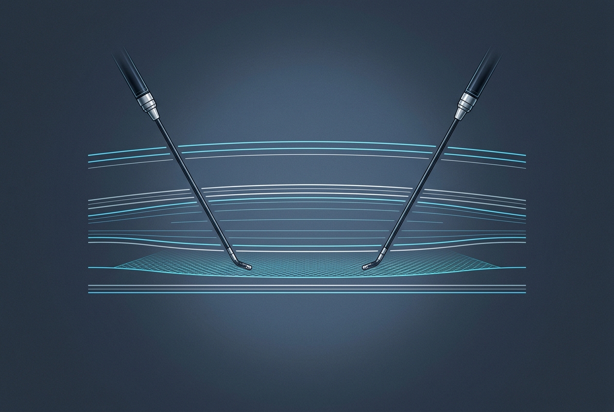
Why sublay, from the front
A sublay mesh sits in a closed space — muscle and anterior fascia above it, posterior fascia, transversalis and peritoneum below — which helps reduce seroma and infection. The detailed, surgeon-level description of the TESAR technique lives on the hub.
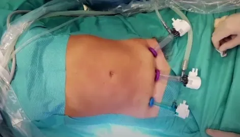
TESAR
Anterior access with the mesh placed sublay, behind the muscle — the technique developed by Dr. Fiori.
Robotic surgery
Robotic surgery is not a new operation but a highly precise way of performing existing minimally invasive techniques. The surgeon sits at a console and operates through robotic arms with 360° articulation. Its advantages are clearest in posterior, retromuscular midline repairs, where ergonomics and suturing are demanding; the robotic versions carry an “r-” prefix (e.g. r-eTEP, r-PeTEP, r-TAPP).
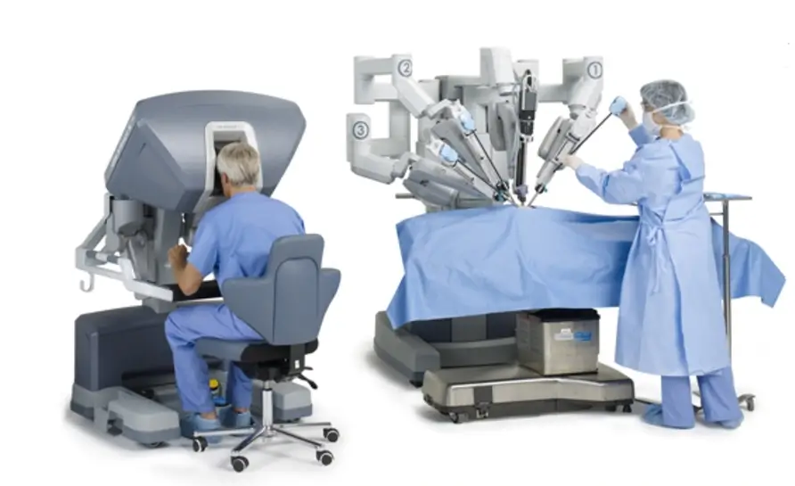
Robotic precision
The robot performs existing minimally invasive repairs with greater ergonomics and precision.
Hybrid techniques
Between open and endoscopic sit the hybrid (video-assisted) techniques: a small incision completed with minimally invasive instruments. Examples are MILOS/eMILOS (a periumbilical access with posterior fascial closure and a retromuscular mesh) and ELAR (a 5–7 cm circumumbilical incision, with the rectus sheaths opened and the muscles approximated over an anterior mesh).
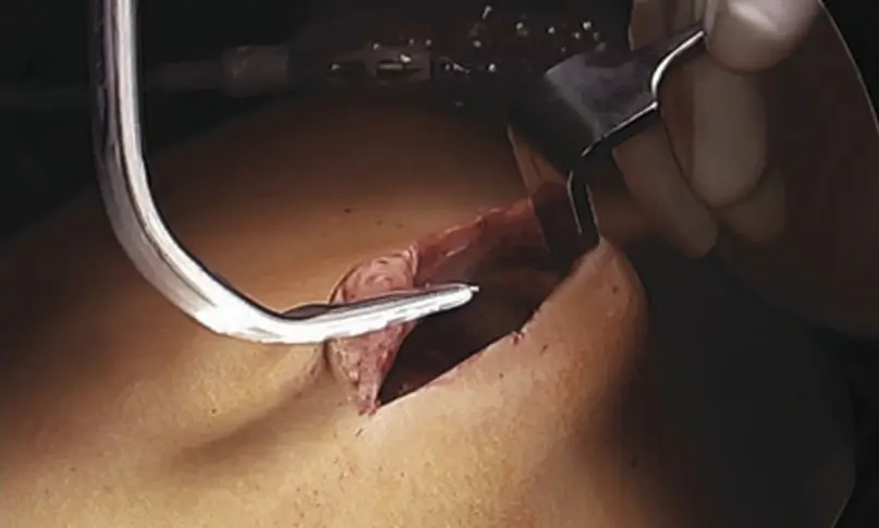
Hybrid techniques
A small incision assisted by minimally invasive instruments, between open and endoscopic surgery.
Where can you see real results?
Rather than duplicating clinical cases here, you can see documented before-and-after results of diastasis surgery, and read how each technique is chosen with a morpho-functional approach, on the hub.
→ See real diastasis results on federicofiori.com. To discuss your own case, the hub explains how to book a consultation.
Documented before/after cases are published only on the surgeon's hub: see diastasis results on federicofiori.com. A photograph documents one person's outcome; the same technique gives a different result on someone else.
When is surgery needed for diastasis recti?
Surgery is proposed when the diastasis causes symptoms — functional and/or morphological. If there are no relevant symptoms it can be deferred, though the wall keeps working inefficiently and symptoms may appear later. There is no single operation for everyone.
Will diastasis surgery give you a flat stomach?
Surgery restores tension and rebuilds a closed, functioning wall, but it does not recreate lost muscle tone, so it does not by itself create a flat stomach. Better pre-operative core tone leads to a more satisfying result.
What is the difference between open and minimally invasive repair?
Open surgery is needed when excess skin and fat must be removed and gives the fullest morphological correction. Minimally invasive repair rebuilds the wall through small incisions but cannot remove major redundant skin.
Who developed the TESAR technique?
TESAR (Totally Endoscopic Sublay Anterior Repair) was developed and first published by Dr. Federico Fiori, in 2019. It uses an anterior endoscopic access with the mesh placed behind the rectus muscle (sublay).