How Rectus Diastasis Is Measured and When It Becomes Significant
Rectus diastasis is measured as the inter-rectus distance, the gap between the two Rectus muscles along the Linea Alba. Manual palpation, ultrasound, and CT are used. A separation is generally considered significant above about 2 to 2.5 cm, though the value alone does not define treatment.
What exactly is being measured in rectus diastasis?
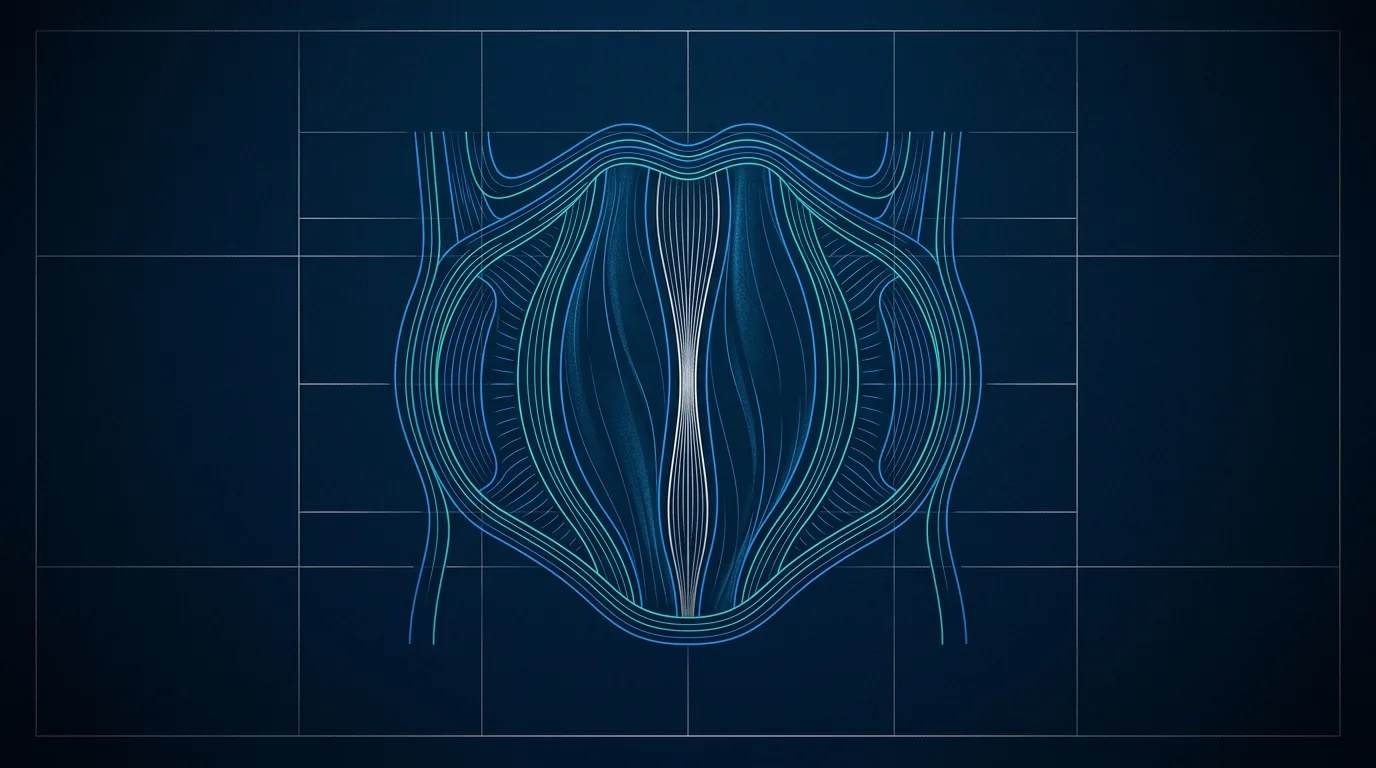
The two Rectus muscles run vertically down the front of the abdomen, one on each side of the midline. Between them lies the Linea Alba, a band of connective tissue that joins them. Diastasis is a widening of this band, so the muscles sit farther apart than usual.
The quantity recorded is the inter-rectus distance, meaning the width of the gap between the medial edges of the two muscles. It is measured with the abdominal wall relaxed and, in some protocols, also during a mild contraction, because the gap can change with effort.
Since the gap is not uniform, the distance is assessed at more than one level: above the navel, at the navel, and below it. Diastasis is often wider around and above the umbilicus, so reporting a single number without its location tells only part of the story.
How is diastasis measured with the fingers?
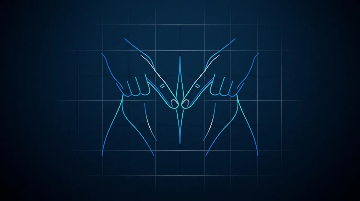
The simplest method is manual palpation. With the Patient lying on the back and knees bent, a gentle lift of the head and shoulders makes the Rectus muscles contract, and the examining fingers sink into the midline gap between them. The number of fingertips that fit gives a rough estimate of the width.
This approach is immediate, needs no equipment, and is useful as a first screening. Its limits, however, are real and should be stated plainly. Finger width varies between examiners, the tension of the wall changes the reading, and abdominal fat makes the edges hard to feel.
For these reasons palpation tends to be operator-dependent and only approximate. It answers the question "is there a diastasis worth looking at?" rather than "exactly how many centimeters is it?". A finding on palpation is generally confirmed with an imaging method before conclusions are drawn.
Why is ultrasound the reference method in most cases?
Ultrasound directly visualizes the two muscle edges and the tissue between them, so the inter-rectus distance can be measured on the screen in millimeters. It is painless, involves no radiation, is widely available, and can be repeated over time to follow changes.
A further advantage is the dynamic study: measurements can be taken both at rest and during a controlled contraction. This helps distinguish a gap that stays open from one that closes on effort, and it also shows whether the Linea Alba bulges outward under pressure.
Ultrasound is nonetheless operator-dependent, since the reading depends on how the probe is held and where the measurement is placed. Standardizing the levels, above, at, and below the navel, and the degree of contraction reduces this variability. In practice ultrasound is considered a reliable reference for most assessments of diastasis.
When is a CT scan needed?
Computed tomography maps the whole abdominal wall in cross-section and is highly reproducible, since it does not depend on the operator's hand. It measures the gap precisely and, importantly, shows the surrounding structures in full.
Its role is not routine. CT involves ionizing radiation, so it is reserved for situations where more information is genuinely needed: a suspected or coexisting hernia, previous abdominal surgery, an unclear clinical picture, or planning of a complex repair where the anatomy must be defined.
For a straightforward assessment of an isolated diastasis, CT is generally excessive. The choice of method follows the clinical question, and ultrasound answers most of them without exposing the Patient to radiation.
From how many centimeters is diastasis considered significant?
There is no single, universally agreed cut-off, and this deserves honesty. The inter-rectus distance is a continuous measurement, and a small gap can be entirely normal, especially in the upper abdomen or after pregnancy in the early months.
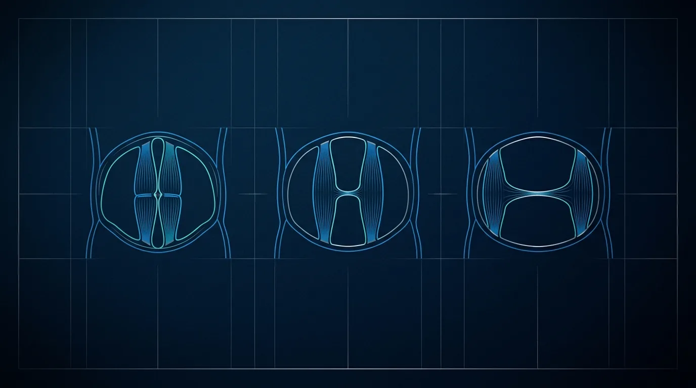
A widely used working threshold treats a distance above roughly 2 to 2.5 cm as beyond the physiological range. Some classifications describe mild, moderate, and larger separations in centimeter bands, but the boundaries differ between sources, so the same measurement may be labeled differently.
The value should therefore be read together with other findings. A gap of a given width means one thing if the tissue is firm and flat, and another if it bulges and causes symptoms. The number opens the discussion; it does not close it.
The elements that weigh alongside the centimeters include:
- the location of the gap along the midline, above or below the navel;
- whether the Linea Alba bulges outward, particularly on effort;
- the presence of symptoms such as discomfort, a sense of core weakness, or functional limitation;
- the coexistence of an umbilical or epigastric hernia, which changes the reasoning entirely;
- the time since pregnancy, since spontaneous improvement continues for several months.
Why the measurement alone does not decide treatment
A common worry is that a certain number of centimeters automatically requires surgery. That is not how the reasoning works. The measurement describes the anatomy; the indication is built on the whole picture, including symptoms, function, and the Patient's own concerns.
In the months following childbirth the wall recovers progressively, and targeted, supervised exercise can improve control and support. For this reason a stable measurement is usually awaited before any surgical discussion, generally not before the postpartum recovery has run its natural course.
When a true hernia coexists with the diastasis, the assessment shifts, because a hernia carries its own indications. Distinguishing a simple widening of the Linea Alba from a hernia with a fascial defect is precisely why imaging matters, and it is one reason the fingers alone are not enough.
How the different methods compare
Each tool answers a slightly different need, and choosing well avoids both under-investigation and unnecessary tests. In brief:
- Palpation: quick, free, no equipment; only approximate and operator-dependent; good for first screening.
- Ultrasound: precise in millimeters, painless, no radiation, can be repeated and done dynamically; still operator-dependent, so protocol matters; suitable as the reference for most cases.
- CT: highly reproducible and complete, shows coexisting hernias and prior surgery; uses radiation and is more costly; reserved for complex or unclear situations.
A sensible pathway usually begins with palpation, is confirmed and quantified with ultrasound, and turns to CT only when the clinical question requires it. Consistency of method over time, same operator where possible and the same measurement levels, makes follow-up figures comparable.
A sober next step
For anyone uncertain about a widening of the abdominal midline, the reasonable move is a clinical evaluation with an ultrasound measurement at defined levels, rather than acting on a single self-measured number. The consultation clarifies where the gap is, whether a hernia coexists, and whether the situation is still evolving after pregnancy. From that complete picture, options can be discussed honestly, including watchful waiting and supervised exercise, without any promise of a fixed result.
Frequently asked questions
- Can rectus diastasis be measured reliably at home?
- A rough self-check is possible by lying down, bending the knees, lifting the head slightly, and feeling the midline gap with the fingers. This gives only an approximate idea and is easily misread, since abdominal fat and muscle tension change the sensation. A reliable, millimeter measurement requires ultrasound performed at defined levels, so a home finding is best confirmed with a clinical assessment rather than acted upon alone.
- Is a 3 cm diastasis always a problem?
- Not necessarily. A distance around 3 cm is generally beyond the physiological range, but the width alone does not define whether treatment is needed. What matters alongside it is the location of the gap, whether the tissue bulges under effort, whether symptoms are present, and whether a hernia coexists. Two people with the same measurement may follow very different paths, decided after a full evaluation.
- Does the gap change when the abdomen is contracted?
- Yes, and this is why a dynamic study is useful. During a mild contraction the Rectus muscles can draw closer, so the gap may narrow, or the Linea Alba may bulge outward if the tissue is weakened. Measuring both at rest and on effort gives a fuller picture than a single resting value and helps distinguish a stable widening from one that behaves poorly under load.
- Should a CT scan be requested to confirm diastasis?
- In most cases it is not necessary. CT is precise and reproducible but uses ionizing radiation, so it is reserved for situations that genuinely need it, such as a suspected coexisting hernia, previous abdominal surgery, an unclear picture, or planning of a complex repair. For an isolated diastasis, ultrasound usually answers the clinical question without radiation exposure, and is the preferred first-line imaging method.
- How long after pregnancy should the measurement be taken?
- The abdominal wall recovers progressively in the months after childbirth, and the gap often narrows on its own during this period. For this reason a stable measurement is generally awaited before drawing conclusions or discussing surgery, rather than measuring in the early weeks. Supervised, targeted exercise during recovery can improve control and support, and a later assessment gives a more meaningful figure to reason from.
People also ask
- What is diastasis recti?
- What are the symptoms of diastasis recti?
- How do you check for diastasis recti at home?
- When does diastasis recti need surgery?
- How is diastasis recti diagnosed?
Pages that cover this topic
- Diastasis recti surgery: before and after, explained
- What is diastasis recti?
- Diastasis recti self-assessment at home
- Diagnosis and imaging of diastasis recti
- Functional anatomy of the abdominal core
- Diastasis recti pictures: how to recognize it at rest and under strain
Published on: 2026-09-07 · Content by Dr. Federico Fiori